New Multimode Scanning X-ray Diffraction Microscopy for Diamond Anvil Cell experiments
Materials subjected to high pressure and high/low temperature conditions in the diamond anvil cell (DAC) often exhibit a large degree of complexity and inhomogeneity, on various length scales ranging from nanometers to hundreds of microns. An ability to perform fast and detailed spatially resolved characterizations of the inhomogeneity has great promise to unlock the complexity and to gain understanding of a multitude of emerging physical phenomena in high pressure sciences.
A new capability has recently been commissioned at HPCAT that utilizes multimode scanning x-ray diffraction microscopy (SXDM) for experimental studies using high pressure devices, such as DAC. The system leverages the hardware-based scanning capability, also recently developed in HPCAT [1]. The new capability uses any measured information, such as diffraction peaks, unit cell dimensions, texture (preferred grain orientations), and anisotropic strain, to construct the corresponding 2D images in a spatially resolved manner. Unlike the traditional single-point x-ray diffraction, the multimode SXDM can now be routinely performed as a full 2D probe, and provides detailed information on phase distribution, morphology, pressure gradient, and sample’s inhomogeneity of the entire region of interest [2].
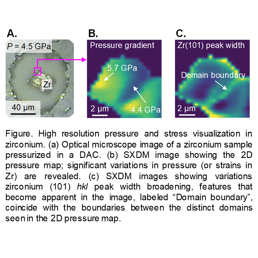
The added value has been briefly demonstrated in case studies on iron and zirconium metal samples under high pressure (See Figure) [2]. These are just a tip of the iceberg; we expect that the true potential of the technique will become apparent on future reports of HPCAT users.
[1] Smith et al., Rev. Sci. Instrum. 90, 015116 (2019)
[2] Hrubiak et al., Rev. Sci. Instrum. 90, 025109 (2019)
Figure. High resolution pressure and stress visualization in zirconium. (a) Optical microscope image of a zirconium sample pressurized in a DAC. (b) SXDM image revealing the 2D pressure map; significant variations in pressure (or strains in Zr) are revealed. (c) SXDM images showing variations zirconium (101) hkl peak width broadening, features that become apparent in the image, labeled “Domain boundary”, coincide with the boundaries between the distinct domains seen in the 2D pressure map.