Synchrotron x-ray shine light on diamond hardness
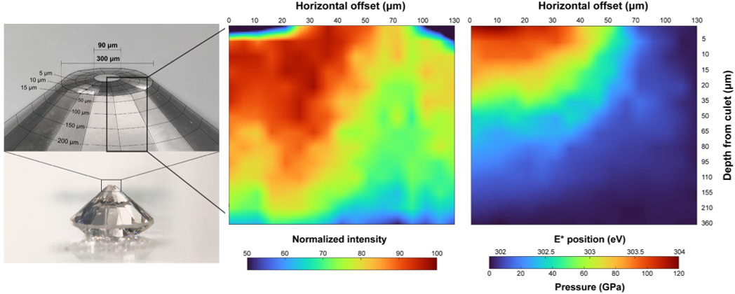
Diamond shows unprecedented hardness. Since hardness is a measure of the resistance to external indentation of chemical bonds in a material, information on the detailed electronic bonding nature of diamond beyond several million atmospheres is key to understanding the origin of hardness. However, probing the electronic structures of diamond at such extreme compression has not been experimentally possible until recently. Researchers used the HPCAT facility and successfully measured the inelastic x-ray-scattering spectra for diamond up to 2 million atmospheres, which provides data on the evolution of its electronic structure under extreme pressure. The mapping of the observed electronic density of states allows us to obtain a two-dimensional image of the electronic bonding transitions of diamond undergoing deformation up to 2 million atmospheres.
The HPCAT results on inelastic x-ray scattering (IXS) unveil the underlying mechanism of diamond’s hardness; its rigidity upon densification is manifested in minor changes in IXS features near the edge-onset for diamond under compression beyond a million atmospheres. Nevertheless, the overall electronic structure exhibits greater flexibility at external pressure, highlighted by the dramatic pressure-induced shift and electron delocalization in full electronic density of state (DOS) pattern, distinct from those observed for other ‘softer’ materials. It is postulated that the observed dispersion likely contributes to the resistance to deform under extreme pressure conditions. Such dual electronic responses, i.e., a minor change in edge-energy onset with flexibility in extended DOS, during compression indicates that external toughness of diamond is supported by its ability to reconcile internal stress. These unique evolutions of electronic structures account for diamond’s unprecedented hardness and rigidity under extreme compression. The information on the pressure-driven changes in the DOS pattern proved by carbon’s K-edge may be useful to explore the electronic origins of hardness and rigidity in diverse materials. The current study highlights the first report of the in-situ two-dimensional image of the electronic bonding transitions with full DOS of any condensed matter, not to mention diamond, undergoing densification above 1 million atmosphere. As IXS is becoming a widely used technique at synchrotron x-ray sources, there is practical significance of proposed protocols, providing a robust pressure-indicator for IXS high-pressure experiments beyond megabar pressure conditions relevant to super-Earth bodies and deep planetary interiors.
For more, see: Sung Keun Lee, Yoosoo Yi, Yong-Hyun Kim, Hyo-im Kim, Paul Chow, Yuming Xiao, Peter Eng, Guoyin Shen, "Imaging of the electronic bonding of diamond at pressures up to 2 million atmospheres," Sci. Advances 9 (20), eadg4159 (2023). DOI: 10.1126/sciadv.adg4159